Laboratory of Cytometry and Image Analysis (CoralWarm)
Keywords: morphology, image, photography, video, microscope, reproductive biology, mesenteric septa, germ cells, embryos
The laboratory of Cytometry and Image Analysis, located in Via Selmi 3, runs research and teaching activities through morphological analyses on aquatic and terrestrial invertebrate organisms. The laboratory is mainly used for reproductive biology studies of cnidarians, through the observation of histological slides and the measurement of germ cells. The laboratory is also used for the observation and manipulation of invertebrates and to perform high-resolution macro photographs.
Laboratory equipment
The Cytometry and Image Analysis laboratory is equipped with:
- 1 Image analysis system consisting of 3 cameras, 4 optical microscopes, 1 macro camera, 2 workstations with dedicated software
- 4 Stereoscopes for observation, preparation, and manipulation of samples
- 1 Macro station for image and video acquisition
- 2 binocular microscopes with digital image acquisition system
Teaching, training, and research activities
The laboratory is available to support the preparation of BSc, MSc, and PhD Theses, and for the implementation of research projects by post-doc fellows and researchers.
Album
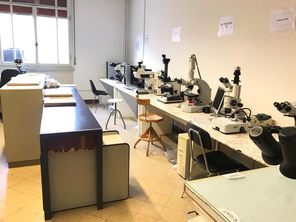
The laboratory of Cytometry and Image Analysis
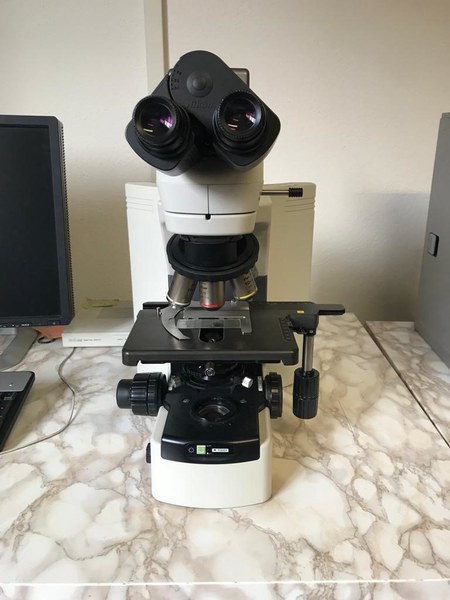
Optical microscope
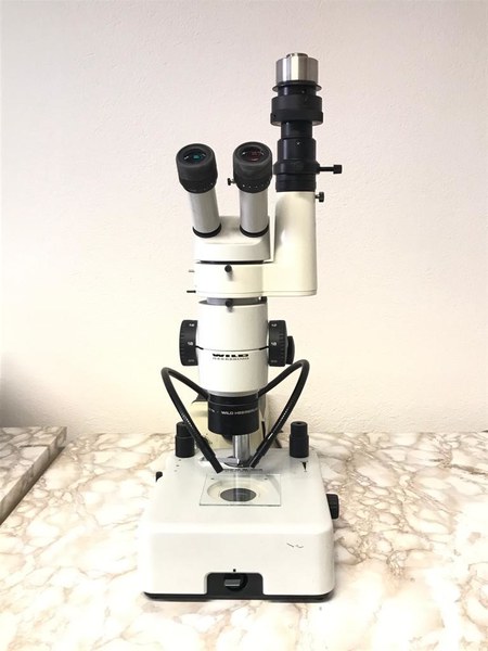
Stereoscope
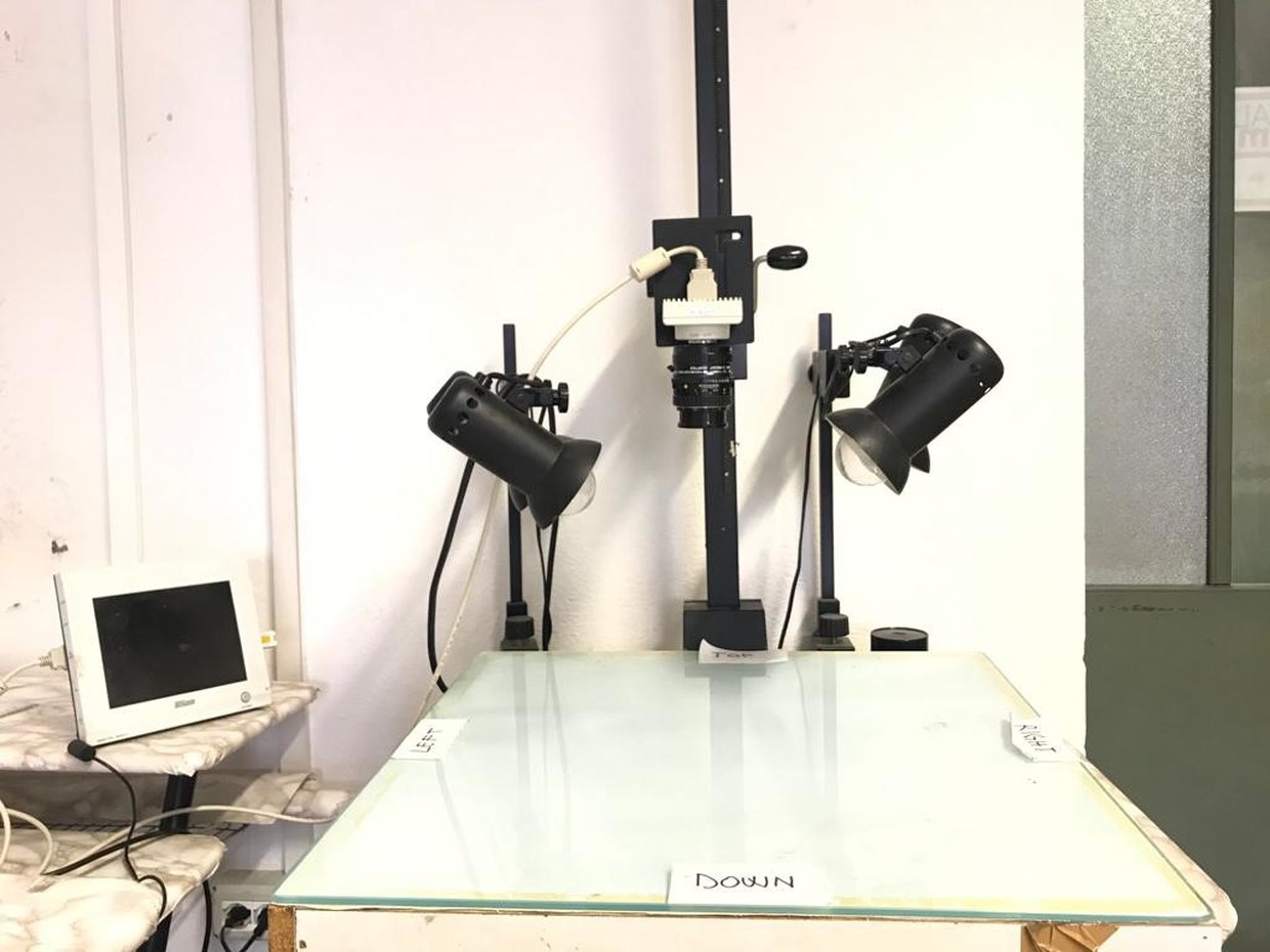
Macro station with camera and monitor
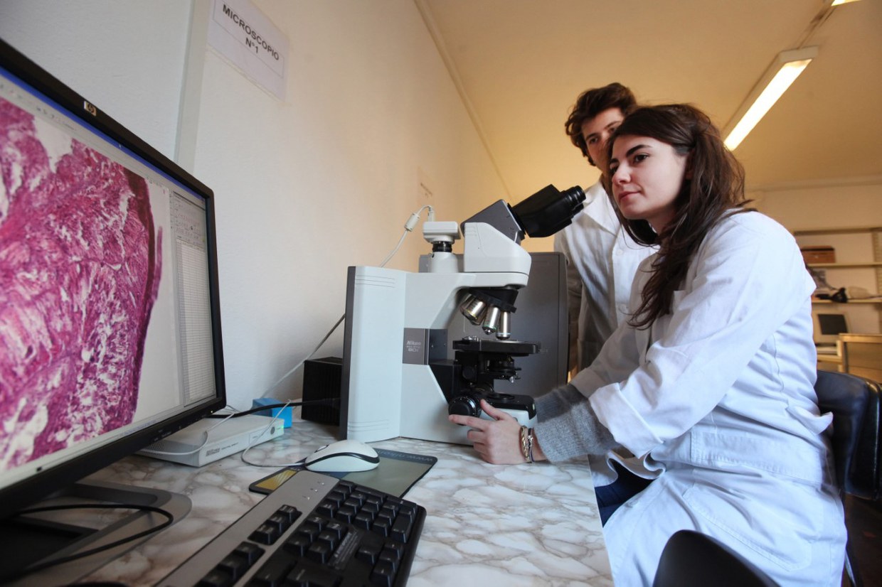
Students looking at histological slides
Contacts
-
Associate Professor
Dipartimento di Scienze Biologiche, Geologiche e Ambientali
Via Selmi 3
Bologna (BO)
Tel: +39 051 20 9 4244
-
Associate Professor
Dipartimento di Scienze Biologiche, Geologiche e Ambientali
Via Selmi 3
Bologna (BO)
Tel: +39 051 20 9 4157
-
Mauro Cesarini
Area dei Funzionari - Settore scientifico - tecnologico
BiGeA - Servizi tecnici di laboratorio Bologna-Fano
Via Selmi 3
Bologna (BO)
Tel: +39 051 20 9 4252
-
Maria Roberta Randi
Area dei Funzionari - Settore scientifico - tecnologico
BiGeA - Servizi tecnici di laboratorio Bologna-Fano
Via Selmi 3
Bologna (BO)
Tel: +39 051 20 9 4209